The Translational Pathology Shared Resource (TPSR) was established in 2011. It provides University of Illinois Cancer Center (UICC) members with access to a comprehensive range of services involving the analysis of animal model and human tissue samples. These services include basic and advanced histological techniques, chemical staining and immunohistochemistry, digital slide scanning, image analysis, tissue microarray design and construction, and laser capture microdissection. In addition, it provides consultation for projects from pathologists with both clinical and laboratory animal expertise.
Our Services
The TPSR supports its users through consultation, direct services, and relevant training and education.
Consultation
The TPSR staff is available to UICC members for consultation regarding:
- Human and laboratory animal pathology
- Histology
- Digital imaging
- Laser capture
- Tissue microarrays (TMAs)
- Patient-derived organoid / xenograft (PDO/PDX) development
Complex projects begin with a free 1-hour consultation that results in a plan and related cost estimate.
For grant proposals, the TPSR provides budget details, text regarding methodology, and information regarding facilities. The TPSR also connects investigators with pathology faculty who have specialized knowledge related to their research.
Direct Services
Histology
Through its Research Histology Core (RHC), TPSR offers a full range of basic histology services, including:
- Specimen processing
- Embedding
- Sectioning
- RNA/DNA extraction
- Direct chemical staining
- Immunohistochemistry
We also provide antibody validation and optimization of immunochemistry protocols.
Advanced histology methods, such as multiplex staining using chromagen or fluorescence labeling, and CISH/FISH are also available.
Imaging
The Research Tissue Imaging Core (RTIC) provides whole-slide digital scanning, allowing users to view, annotate, and share histology images with free Web-based software (Aperio ImageScope®).
Projects requiring image analysis begin with a consultation involving both histology and imaging specialists in order to ensure that staining is optimal.
A suite of image analysis modules from Indicalabs (HALO®) serves as the main platform for most brightfield (and selected fluorescence) analyses. These modules include machine learning capability and algorithms for a wide range of digital pathology tasks, including:
- Quantification of signals in various cell compartments
- Area quantification (e.g., inflammatory foci)
- Tumor vascularization analysis
- Spatial analysis
- ISH
- TMA analysis
- Serial section registration
Images obtained on the multispectral Vectra system are typically analyzed using inForm® and Phenoptics® software (Akoya Biosciences), whose capabilities include spatial analysis of multiple molecular or cellular targets in an image. A Nanostring nCounter® instrument is available for image-based quantification of multiplexed, bar-coded RNA transcripts from tissue or cell specimens.
TMA Services
To receive TMA services, UICC members meet with a research pathologist (Dr. V. Macias) to design the TMA. Once IRB approval is obtained and a design is final, archival tissue blocks are accessioned, usually through the UI Biorepository. Macias uses whole-slide scans of H&E-stained sections to select areas for core punching using either an automated or manual array device.
Patient-Derived Models
Investigators interested in PDX/PDO models either receive training in how to develop and maintain models in their own labs or direct services involving the entire process, from harvesting of primary cells to initial culturing and propagation/expansion of specimens in either xenografts or 3D culture.
Training & Education
The TPSR provides education and training on:
- Preparation of tissue samples for histological analysis, including microtome/cryostat use
- Laser capture microdissection
- Basic image analysis
- Developing and maintaining PDX and PDO specimens
TPSR organized three seminars in 2019-2020, co-sponsored with Nanostring and Akoya focusing on multiplex spatial imaging.
Meet Our Team

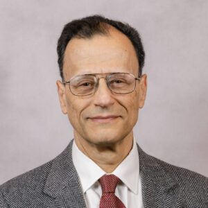
Andre Kajdacsy Balla, MD, PhD, TPSR Leader

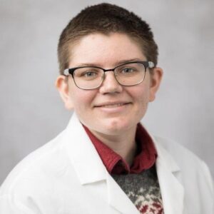
Eileen Brister, PhD, Research Tissue Imaging Core

Subhasis Das, MSc, PhD, Patient-Derived Models Laboratory

Maria Sverdlov, PhD, MS, Research Histology


