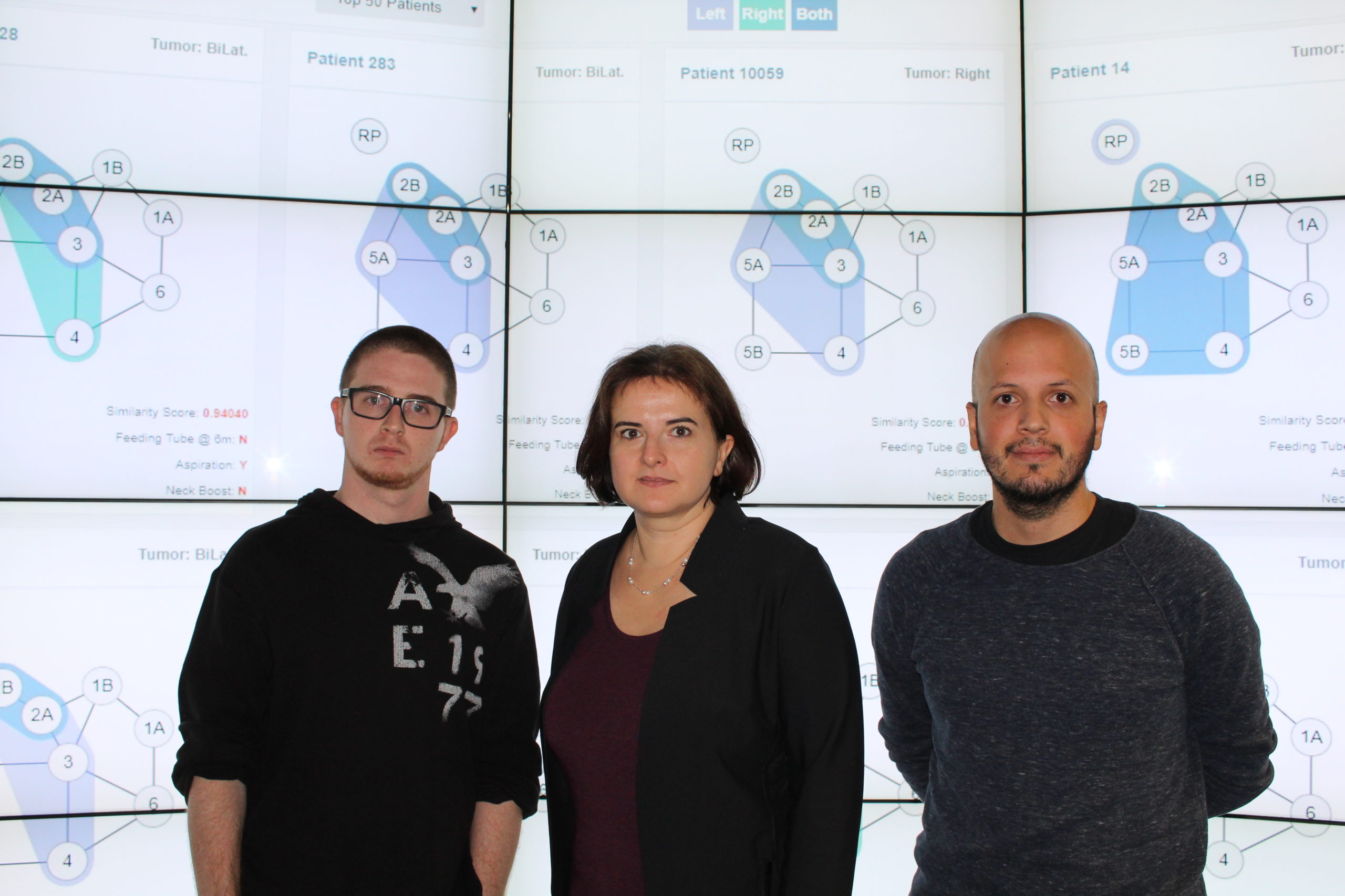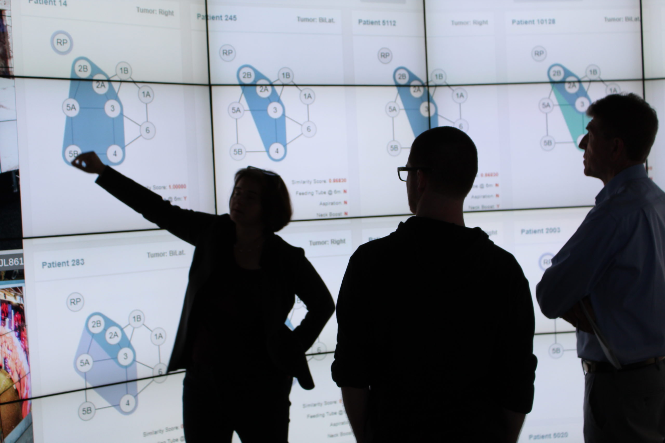
Computer applications come in a variety of forms: computer vision, autonomous vehicles, text mining, health monitoring mobile apps. G. Elisabeta Marai is taking it further. She is designing computer programs that will assist medical professionals in determining the best treatment options for cancer patients, specifically those suffering from head and neck cancer.

As associate professor of computer science at UIC’s Electronic Visualization Laboratory, Marai’s research focuses on visual computing, an area of computer science that handles computing with images and 3D models, as well as the processes that happen at the interface between humans and data that can be represented visually. Prior to being introduced to an oncology radiologist at Houston’s MD Anderson Cancer Center a few years ago, Marai hadn’t thought about combining her expertise in computer science with cancer, beyond working with bioinformatics cell signaling data. But the conversation intrigued her about how the two could collaborate.
“Head and neck cancer can be treated in multiple ways, from radiation alone to a combination of radiation and chemotherapy or induction chemotherapy. But the one thing that’s common is they rely on imaging to develop treatment options,” said Marai, who is also a member of the University of Illinois Cancer Center. “These decisions depend on complex factors, including the tumor location in relation to sensitive organs and its response to treatment, laboratory data, toxicity, anticipated side effects and survival probability.”
Marai and her collaborators at MD Anderson Cancer Center, University of Iowa, and University of Minnesota obtained a R01 grant (CA214825-01) from the National Institutes of Health (NIH) and National Cancer Institute (NCI) to develop a computing methodology that will precisely determine health outcomes for patients with head and neck cancers based on demographics, toxicity and complex imaging data. R01, or Research Project Grant, is the original and historically oldest grant mechanism used by NIH. It provides support for health-related research and development based on the organization’s mission.
 Head and neck cancers affect all ethnicities and age groups, but they are more than twice as common in men as women. Correlated with the wider spread of HPV, the number of such cancers is increasing. At least 75 percent of head and neck cancers are, furthermore, caused by tobacco and alcohol use. More than 50,000 new cases are diagnosed each year in the United States, leading to “large, rich repositories of patient data,” Marai said.
“To select the best treatment option that balances efficacy and toxicity, oncologists need to anticipate survival, oncologic, and side effect outcomes,” she said. “However, despite the wealth of available data, risk prediction algorithms for cancers are rudimentary and incorporate minimal patient characteristics, largely due to a lack of computational methodologies.”
The program will be the first to include both complex imaging and non-imaging data, while utilizing large-scale biological and clinical variables. It is unique, Marai said, because it combines principles from bioengineering, statistics and computer science.
“Our model and web-based environment will mark a significant breakthrough in biomedical computing because it will be able to identify, for the first time, specific subgroups who are at-risk for distinct oncologic, toxicity and survival profiles,” Marai said. “The current clinical practice paradigm for head and neck treatment is stage-driven. Our approach can identify patients based not on clinical staging, but on precision modeling using cohort data and similar sets of patients.
“This has the potential to change the standard of care and outcomes of treatment.”
Through a second grant obtained from the NIH and the NCI (R01CA225190-01), Marai and her colleagues and students, including graduate students Tim Luciani, Juan Trelles and Peter Hanula, are developing computational models that combine imaging data, such as radiation dosage and spatial distribution, and nonspatial data, such as demographics and toxicity. Data derived from the computer models will identify similar patients and assist medical professionals in predicting the optimal treatment options.
The computer program has the potential to improve the standard of care – a treatment plan selected by the tumor board – and the quality of life of surviving patients, Marai said. It can also be used to develop the best treatment strategies not only in head and neck, lung, or brain cancer, but also other chronic conditions such as mental health disorders, substance abuse diseases, or diabetes, illnesses that require making multiple decisions that adjust for efficacy and toxicity.
Marai began combining computer science with medicine while in graduate school at Brown University. Under the guidance of David Laidlaw, PhD, professor of computer science and Joseph “Trey” Crisco, PhD, professor of orthopaedics and engineering, Marai conducted research in visual computing for biological applications, working with sequences of computed tomography (CT) volumes to extract articulation motion and to computationally model soft tissues. She created novel computational modeling, visualization and analysis tools that are needed to model anatomical joints and their variation with disease progression. Following graduation from the Providence, R.I., school, she continued working with orthopaedics data, and expanded her interests into other biomedical fields, including epidemiology and biology.
